

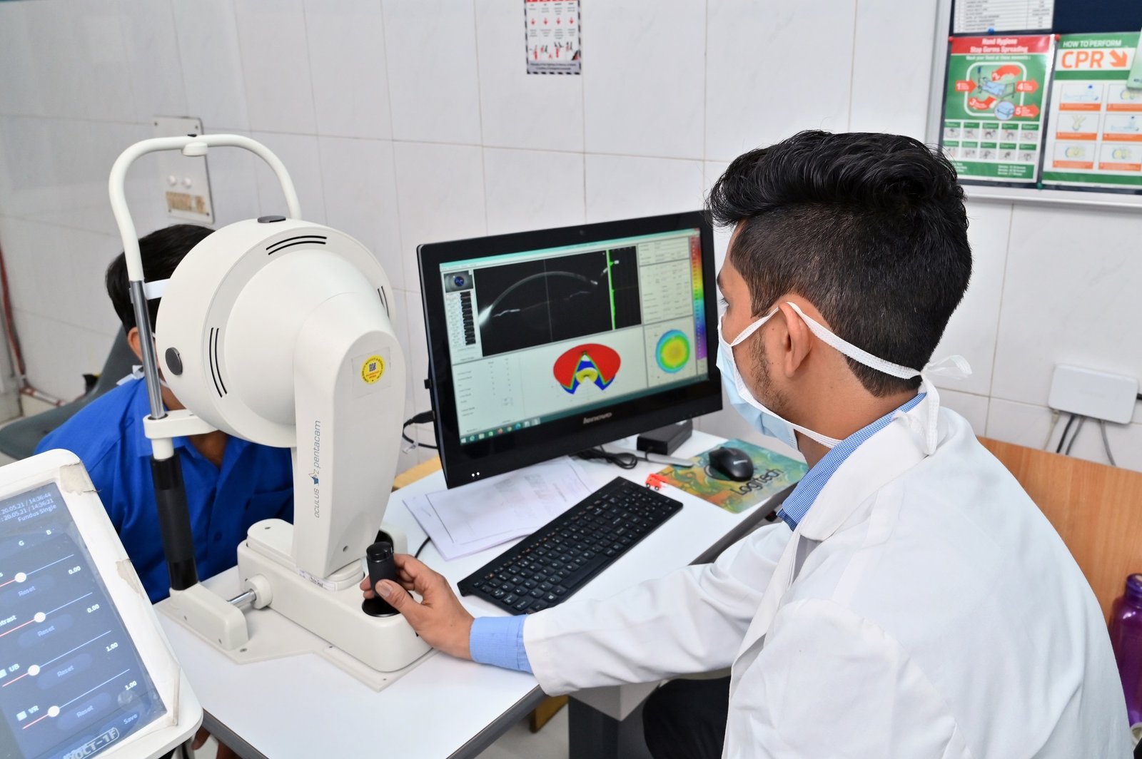
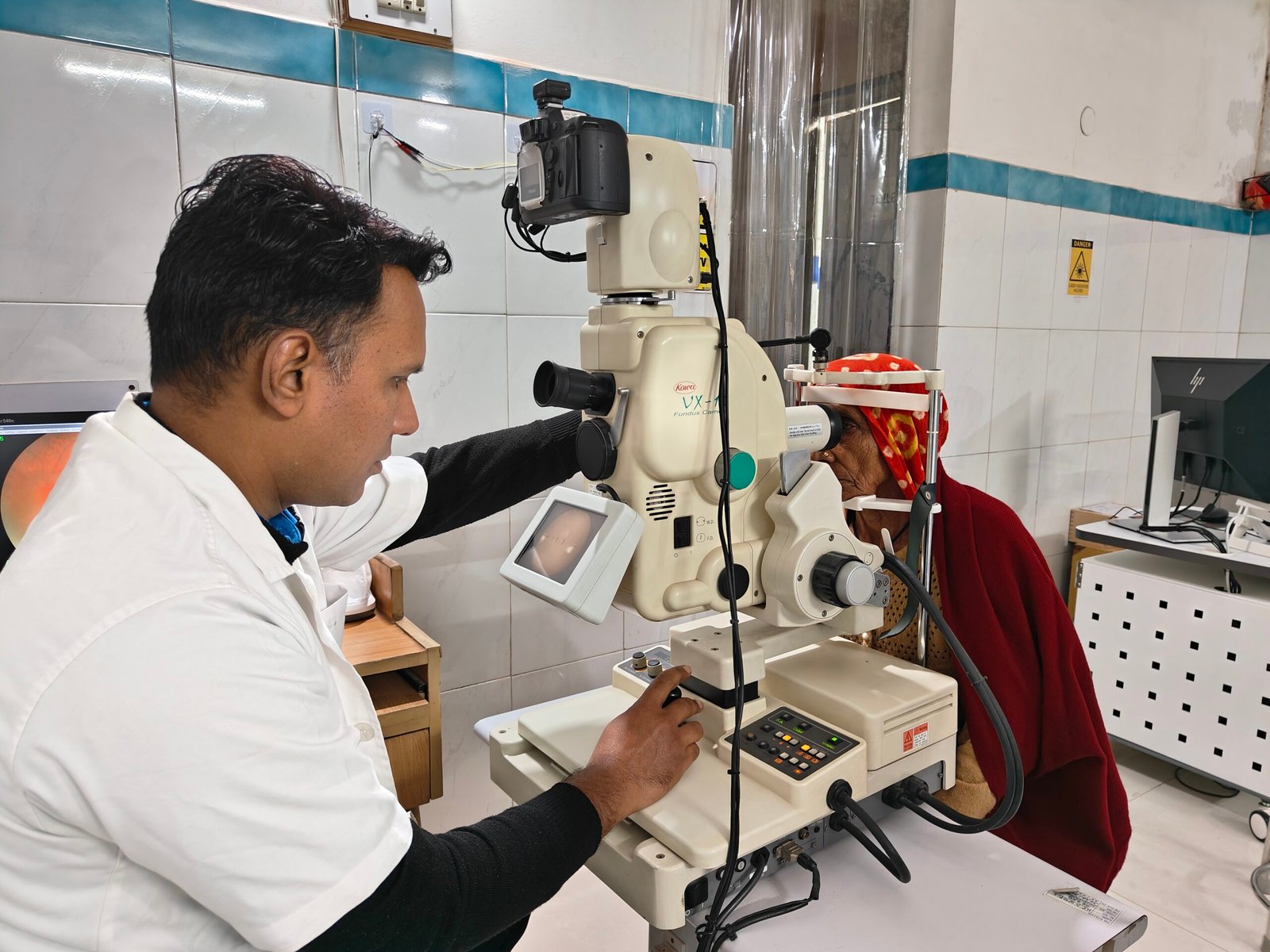
At Dhir Hospital, we have a dedicated team of cornea specialists committed to provide you with the best possible care to protect your vision.
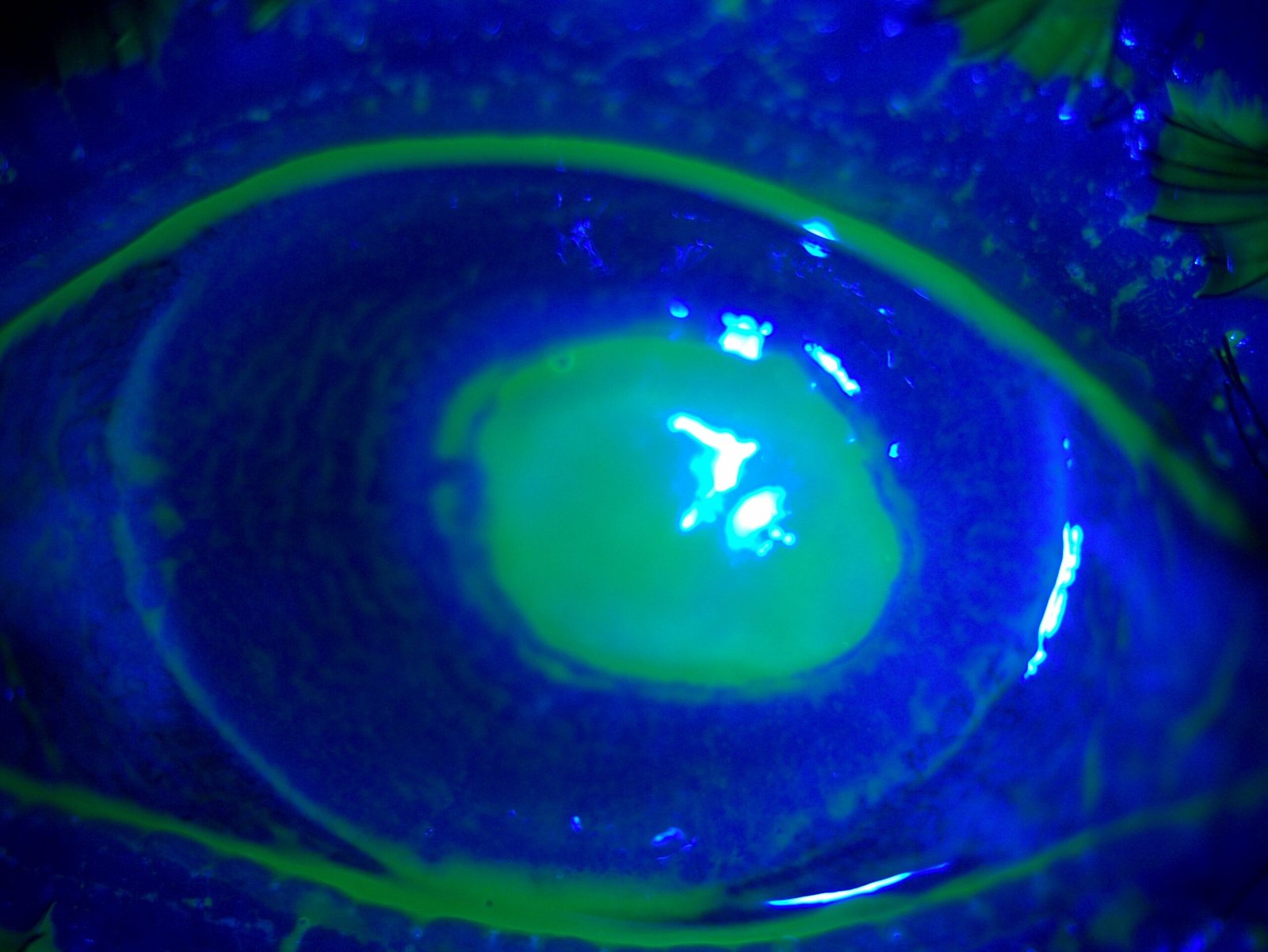
The cornea is like the clear front window of your eye, helping you see by focusing light. It’s crucial for clear vision, but it can affect how well you know if it gets damaged or cloudy. Glasses or contact lenses can fix minor issues, but a cornea transplant may be needed in severe cases. During a transplant, it’s like giving your eye a new clear window. A surgeon replaces the damaged cornea with a healthy one from a donor. It’s like getting a new piece of glass for your eye, improving vision and reducing problems like blurry vision. This safe and common procedure can greatly affect how well you can see.
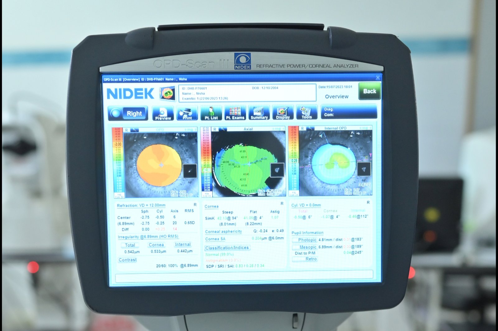
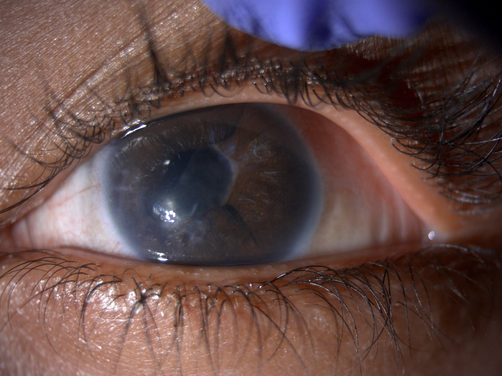
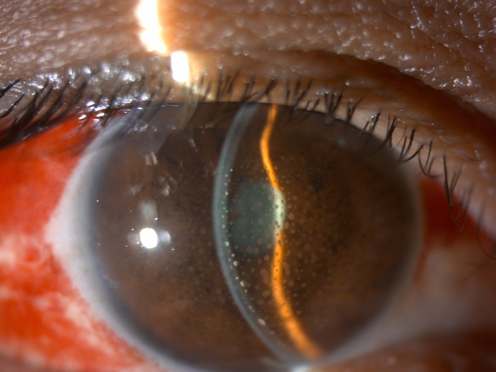
A cornea transplant surgery, done by cornea specialists, is often performed to help people see better when their cornea is damaged. It can also ease pain and symptoms linked to cornea diseases. Various conditions like a bulging cornea (keratoconus), a genetic issue called Fuchs dystrophy, thinning or tearing of the cornea, scarring from infection or injury, swelling, corneal ulcers not responding to medicine, and problems from past eye surgeries can be treated with corneal transplants.
A cornea transplant is quite safe, but there is a slight chance of serious issues, including
Corneas for transplants come from individuals who have passed away. However, corneas from those with unknown causes of death, a history of eye surgery, eye disease, or certain contagious conditions are not used. Unlike other organ transplants, there is no need for tissue matching for cornea recipients. There is usually no lengthy waiting list for cornea transplants in the United States, as donor corneas are readily available.
A cornea transplant entails the removal of either the complete or partial thickness of a diseased cornea, substituting it with healthy donor tissue. The choice of method is determined by the cornea surgeon based on individual circumstances. These procedures include:
Penetrating keratoplasty, also known as full-thickness cornea transplantation, has been used for over a century and remains a valuable procedure. During this surgery, the surgeon uses a tiny circular blade to remove the entire center of the damaged cornea. Subsequently, it is replaced with a healthy piece of donor cornea that matches the same shape. This helps restore vision and has benefited thousands of individuals each year.
Endothelial keratoplasty involves two types of operations that target removing diseased tissue from the back layers of the cornea, including the endothelium and the Descemet membrane attached to it. In both procedures, donor tissue is utilized to replace the removed tissue.
The first operation, Descemet stripping endothelial keratoplasty (DSEK), employs donor tissue to replace up to one-third of the cornea.
The second operation, Descemet membrane endothelial keratoplasty (DMEK), uses an even thinner and fragile layer of donor tissue. While more challenging than DSEK, DMEK is a commonly used procedure.
Deep anterior lamellar keratoplasty, or partial thickness cornea transplant, is a procedure conducted when the innermost layer of your cornea is healthy. Still, damage is present in the middle and outer layers. In this surgery, your surgeon removes the affected middle and outer layers of the cornea, replacing them with healthy donor corneal tissue. This helps restore the cornea’s function and improve vision.
What can you expect
After the Procedure
After a cornea transplant, many individuals experience at least partial vision restoration. The post-transplant outcomes depend on your overall health and the purpose of the surgery.
Ongoing annual checkups with your eye doctor are crucial to monitor for potential complications and cornea rejection, which can persist for years. Fortunately, medications are often effective in managing cornea rejection.
Following cornea transplant surgery, your vision may initially worsen, requiring time for adjustment to the new cornea. The healing process of the outer corneal layer may take weeks to months, during which your cornea specialist will make necessary adjustments to enhance vision. These adjustments include correcting irregularities caused by stitches holding the donor cornea, addressing astigmatism, and managing refractive errors like near-sightedness and farsightedness. Your cornea specialist may recommend glasses, contact lenses, or, in some cases, laser eye surgery for optimal vision correction. Regular check-ups with your cornea specialist are important.

Dhir Hospital has the best Cataract & Anterior Segment in Bhiwani, Haryana with decades of experience.
At Dhir Hospital, we provide the best Retina & ROP Treatment in Bhiwani, Haryana with experienced Retina Surgeons.
At Dhir Hospital, we provide the best Glaucoma Treatment in Bhiwani, Haryana with experienced Glaucoma Surgeons.
At Dhir Hospital , we are committed to providing our uveitis patients with advanced quality eye care at affordable prices.
A corneal transplant, also known as a corneal graft or keratoplasty, is a surgical procedure that involves replacing a damaged or diseased cornea with a healthy cornea from a donor. The cornea is the clear, dome-shaped tissue at the front of the eye that plays a crucial role in focusing light onto the retina for clear vision.
At Dhir Hospital in Bhiwani, Haryana, we are proud to have our own dedicated eye bank, which plays a vital role in providing corneal tissue for transplants. Dhir Hospital Eye Bank is responsible for the meticulous collection, evaluation, preservation, and distribution of corneal tissue from deceased donors. This ensures a reliable and readily available supply of high-quality donor corneas for our patients in need of corneal transplants.
By having our own eye bank, we have greater control over the quality and availability of corneal tissue, allowing us to offer timely corneal transplants to our patients in need. At Dhir Hospital, we are committed to providing the highest standard of care and ensuring the best possible outcomes for corneal transplant recipients.
The eye is a remarkable and intricate organ that allows us to perceive the world around us. Let us explain how it works:
In addition to these essential components, other structures within the eye, such as the aqueous and vitreous humor, help maintain the shape and nourishment of the eye. The muscles surrounding the eye allow for its movement and proper alignment.
Treatment options for corneal conditions vary depending on the specific condition but may include medication (such as eye drops or ointments), contact lenses, corneal transplant surgery, or other surgical interventions.
Designed by Digital Amigos, Maintained by RS Technology